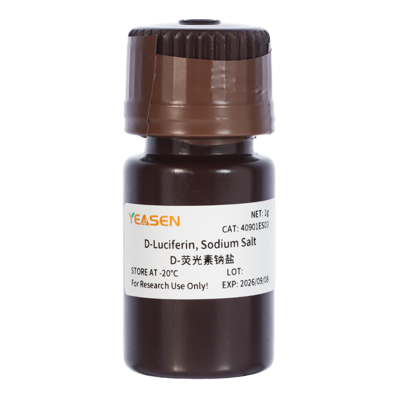
D-Luciferin, Sodium Salt D 荧光素钠盐
勾选需要的规格可一次提交;也可以逐个询价。货号会一并带入询价单。
Q:荧光虫荧光素( Firefly Luciferin)、甲虫荧光素( Beetle Luciferin)和 D-Luciferin 有区别吗?
A:无区别。三者仅仅是不同公司在命名上的差异,均是化合物 (S)-2-(6-Hydroxy-2-benzothiazolyl)-2
-thiazoline-4-carboxylic acid。
Q:该系列产品主要用于哪些应用?
A:除用于活体成像外,荧光素类产品还用于荧光素酶参与的其他应用中,如 体外报告基因检测、微生物/病毒监测、焦磷酸测序等
Q:荧光素钠盐和 D-荧光素钠盐的区别?
A:荧光素钠盐:可用于细胞通透性检测,通过检测 OD490 处吸光度值测定 颜料的通透性。 D-
荧光素钠盐:用于活体成像和报告基因系统,通过冷发光模块检测,不 需要激发光激发。
Q:荧光素钾盐、钠盐、自由酸区别?
A:三者的区别主要在于: 1)溶解性:盐形式易溶于水,其中钾盐溶解度为 60mg/ml,钠盐溶解度为 100mg/ml。自由酸不易溶于水,可用碳酸氢钠溶液弱碱调节溶解,其 在甲醇中的溶解度为10mg/ml,DMSO 中溶解度为 50mg/ml。 2)毒性方面:盐形式在使用过程中较方便,尤其是在体内成像实验中, 因其能够溶解在水中,反应毒性也会更小些(荧光素是一种由苯丙噻唑和 噻唑羧酸基团组成的低分子量有机化合物,有低毒性)。3)使用效果:无明显差异。在体内实验研究中,选用钾盐使用率较高。
Q:荧光素的纯度对实验有成影响吗?
A:有影响。99%以上的纯度较好。对于 99%纯度的荧光素,有 1%的固体杂质。若是这 1 g 的杂质溶解在25ml 的缓冲液中(该稀释比例是进行活体成像实验的标准稀释方法),此时杂质的浓度为 0.4 g/L。假 设杂质的分子量为 1000 g/mol,那么杂质的物质的量浓度为 400 μM,该浓度可能会抑制细胞内某些酶的作 用,并且有可能降低实验效果或对动物产生伤害。
Q:产品稳定性如何?
A:粉末避光保存于-20 或-70℃,有效期至少 1 年。
Q:活体成像底物作用原理?
A:D-荧光素(D-Luciferin)是荧光素酶(Luciferase)的常用底物。其作用机制是在 ATP 和荧光素酶的作用下,荧光素(底物)能够被氧化发光。当荧光素过量时,产生的光量子数与荧光素酶的浓度呈正相关性。2、天然腔肠素(Coelenterazine native)是海肾荧光素酶(Rluc)和 Gaussia 荧光素酶(Gluc) 等多种荧光素酶的作用底物。
Q:推荐仪器?多功能酶标仪能用吗?
A:推荐仪器:1、具有生物化学发光检测模块。荧光素产生的光可以被光度计或闪烁计数器检测。常见的活体成像仪器:如 IVIS® Lumina 小动物活体成像系统,德国 Bruker 公司的 In-Vivo Xtreme 多模式小动物活体成像仪。2、多功能酶标仪:需要和仪器厂家确认是否具体生物化学发光检测模块(注: 不能用荧光显微镜。)
Q:底物的激发波长和发射波长?
A:化学发光,无需激发波长。萤火虫荧光素发射波长是 560nm。海肾荧光素发射波长是 465 nm。
Q:底物可以进入活细胞中吗?
A:可以通过血脑屏障,胎盘屏障和血液测试屏障,也可以进入活细胞。
Q:荧光素类注射方式和用量?
A:下面建议浓度来源于文献:1)注射方式:可通过腹腔注射或尾静脉注射 2)注射量:科学的方法是根据动力学曲线评估注射剂量。建立最初尝试注射剂量为:150mg 底物/kg 小鼠体重。因此,购买量可按照上述方法计算:若 10 只小鼠,22 -25g,则需要底物 33 -37.5mg 。
Q:荧光素的发光特性如何?
A:荧光素腹腔注射老鼠后约 3 min 后,能够表达荧光素酶的细胞开始发光,10 min 后强度达到稳定的最高点,在最高点持续约 10 - 15 min 后开始衰减,可在注射后 10 -15 min 内检测。仅供参考,建议预实验建立荧光素酶 动力学曲线,从而确定最高信号检测时间和信号平台期
Q:活体成像实验,无效果的原因?
A:成功进行活体成像实验需要以下条件:目的组织或细胞表达荧光素酶基因;荧光素底物注射成功; 依赖于发光部位的组织厚度。若实验不成功,可从上述因素查找,荧光素酶基因是否表达,荧光素底物是否未正确注射,及发光部位较深等
已发表文献
[1] Guo Y, Guo Y, Chen C, et al. Circ3823 contributes to growth, metastasis and angiogenesis of colorectal cancer: involvement of miR-30c-5p/TCF7 axis. Mol Cancer. 2021;20(1):93. Published 2021 Jun 25. doi:10.1186/s12943-021-01372-0(IF:27.401)
[2] Wang Z, Yu L, Wang Y, et al. Dynamic Adjust of Non-Radiative and Radiative Attenuation of AIE Molecules Reinforces NIR-II Imaging Mediated Photothermal Therapy and Immunotherapy. Adv Sci (Weinh). 2022;9(8):e2104793. doi:10.1002/advs.202104793(IF:16.806)
[3] Liu Q, Sheng Z, Cheng C, et al. Anesthetic Propofol Promotes Tumor Metastasis in Lungs via GABAA R-Dependent TRIM21 Modulation of Src Expression. Adv Sci (Weinh). 2021;8(18):e2102079. doi:10.1002/advs.202102079(IF:16.806)
[4] Zhou M, Zhang J, Shen J, et al. Hydrogen sulfide-linked persulfidation of ABI4 controls ABA responses through the transactivation of MAPKKK18 in Arabidopsis. Mol Plant. 2021;14(6):921-936. doi:10.1016/j.molp.2021.03.007(IF:13.164)
[5] Wang J, Wang C, Li Y, et al. Potential of peptide-engineered exosomes with overexpressed miR-92b-3p in anti-angiogenic therapy of ovarian cancer. Clin Transl Med. 2021;11(5):e425. doi:10.1002/ctm2.425(IF:11.492)
[6] Mao Z, Wei X, Li L, et al. Arabidopsis cryptochrome 1 controls photomorphogenesis through regulation of H2A.Z deposition. Plant Cell. 2021;33(6):1961-1979. doi:10.1093/plcell/koab091(IF:11.277)
[7] Xu P, Chen H, Li T, et al. Blue light-dependent interactions of CRY1 with GID1 and DELLA proteins regulate gibberellin signaling and photomorphogenesis in Arabidopsis. Plant Cell. 2021;33(7):2375-2394. doi:10.1093/plcell/koab124(IF:11.277)
[8] Lin S, Wen Z, Li S, et al. LncRNA Neat1 promotes the macrophage inflammatory response and acts as a therapeutic target in titanium particle-induced osteolysis. Acta Biomater. 2022;142:345-360. doi:10.1016/j.actbio.2022.02.007(IF:8.947)
[9] Zhang Z, Zeng D, Zhang W, et al. Modulation of oxidative phosphorylation augments antineoplastic activity of mitotic aurora kinase inhibition. Cell Death Dis. 2021;12(10):893. Published 2021 Sep 30. doi:10.1038/s41419-021-04190-w(IF:8.469)
[10] Wang L, Tong X, Zhou Z, et al. Circular RNA hsa_circ_0008305 (circPTK2) inhibits TGF-β-induced epithelial-mesenchymal transition and metastasis by controlling TIF1γ in non-small cell lung cancer. Mol Cancer. 2018;17(1):140. Published 2018 Sep 27. doi:10.1186/s12943-018-0889-7(IF:7.776)
[11] Cao X, Xu P, Liu Y, et al. Arabidopsis cryptochrome 1 promotes stomatal development through repression of AGB1 inhibition of SPEECHLESS DNA-binding activity. J Integr Plant Biol. 2021;63(11):1967-1981. doi:10.1111/jipb.13168(IF:7.061)
[12] Du SS, Li L, Li L, et al. Photoexcited Cryptochrome2 Interacts Directly with TOE1 and TOE2 in Flowering Regulation. Plant Physiol. 2020;184(1):487-505. doi:10.1104/pp.20.00486(IF:6.902)
[13] Zhou H, Qi Z, Pei P, et al. Biocompatible nanomicelles for sensitive detection and photodynamic therapy of early-stage cancer. Biomater Sci. 2021;9(18):6227-6235. Published 2021 Sep 14. doi:10.1039/d1bm00847a(IF:6.843)
[14] Zhang L, Zhou J, Yan Y, et al. Excipient-free nanodispersion of 7-ethyl-10-hydroxycamptothecin exerts potent therapeutic effects against pancreatic cancer cell lines and patient-derived xenografts. Cancer Lett. 2019;465:36-44. doi:10.1016/j.canlet.2019.08.019(IF:6.508)
[15] Geng Y, Duan H, Xu L, et al. BMP-2 and VEGF-A modRNAs in collagen scaffold synergistically drive bone repair through osteogenic and angiogenic pathways. Commun Biol. 2021;4(1):82. Published 2021 Jan 19. doi:10.1038/s42003-020-01606-9(IF:6.268)
[16] Qiu J , Peng P , Xin M , et al. ZBTB20-mediated titanium particle-induced peri-implant osteolysis by promoting macrophage inflammatory responses. Biomater Sci. 2020;8(11):3147-3163. doi:10.1039/d0bm00147c(IF:6.183)
[17] Zhou C, Wang G, Jing W, Tan X, Guo H. Anticancer Properties and Mechanisms of Singly-Protonated Dehydronorcantharidin Silver Coordination Polymer in a Bladder Cancer Model. Front Pharmacol. 2021;12:618668. Published 2021 Feb 23. doi:10.3389/fphar.2021.618668(IF:5.811)
[18] Sheng Y, Yu H, Pan H, et al. Genome-Wide Analysis of the Gene Structure, Expression and Protein Interactions of the Peach (Prunus persica) TIFY Gene Family. Front Plant Sci. 2022;13:792802. Published 2022 Feb 17. doi:10.3389/fpls.2022.792802(IF:5.754)
[19] Jia MZ, Liu LY, Geng C, Jiang J. Activation of 1-Aminocyclopropane-1-Carboxylic Acid Synthases Sets Stomatal Density and Clustered Ratio on Leaf Epidermis of Arabidopsis in Response to Drought. Front Plant Sci. 2021;12:758785. Published 2021 Dec 6. doi:10.3389/fpls.2021.758785(IF:5.754)
[20] Xuan Z, Zhao L, Li Z, et al. EPS8L3 promotes hepatocellular carcinoma proliferation and metastasis by modulating EGFR dimerization and internalization. Am J Cancer Res. 2020;10(1):60-77. Published 2020 Jan 1. (IF:5.177)
[21] Chen G, Fan XY, Zheng XP, Jin YL, Liu Y, Liu SC. Human umbilical cord-derived mesenchymal stem cells ameliorate insulin resistance via PTEN-mediated crosstalk between the PI3K/Akt and Erk/MAPKs signaling pathways in the skeletal muscles of db/db mice. Stem Cell Res Ther. 2020;11(1):401. Published 2020 Sep 16. doi:10.1186/s13287-020-01865-7(IF:5.116)
[22] Chen G, Fan XY, Zheng XP, Jin YL, Liu Y, Liu SC. Human umbilical cord-derived mesenchymal stem cells ameliorate insulin resistance via PTEN-mediated crosstalk between the PI3K/Akt and Erk/MAPKs signaling pathways in the skeletal muscles of db/db mice. Stem Cell Res Ther. 2020;11(1):401. Published 2020 Sep 16. doi:10.1186/s13287-020-01865-7(IF:5.116)
[23] You D, Du D, Zhao X, Li X, Ying M, Hu X. Mitochondrial malic enzyme 2 promotes breast cancer metastasis via stabilizing HIF-1α under hypoxia. Chin J Cancer Res. 2021;33(3):308-322. doi:10.21147/j.issn.1000-9604.2021.03.03(IF:5.087)
[24] Qin L, Zhao R, Chen D, et al. Chimeric antigen receptor T cells targeting PD-L1 suppress tumor growth. Biomark Res. 2020;8:19. Published 2020 Jun 3. doi:10.1186/s40364-020-00198-0(IF:4.866)
[25] Zuo X, Xiang W, Li K, et al. MdGRF11, a growth-regulating factor, participates in the regulation of flowering time and interacts with MdTFL1/MdFT1 in apple. Plant Sci. 2022;321:111339. doi:10.1016/j.plantsci.2022.111339(IF:4.729)
[26] Yang X, Yu Q, Xu H, Zhou J. Upregulation of CD22 by Chidamide promotes CAR T cells functionality. Sci Rep. 2021;11(1):20637. Published 2021 Oct 19. doi:10.1038/s41598-021-00227-4(IF:4.380)
[27] Mei Z, Zhang K, Lam AK, et al. MUC1 as a target for CAR-T therapy in head and neck squamous cell carinoma. Cancer Med. 2020;9(2):640-652. doi:10.1002/cam4.2733(IF:3.357)
[28] Zhang Z, Liu L, Cao S, Zhu Y, Mei Q. Gene delivery of TIPE2 inhibits breast cancer development and metastasis via CD8+ T and NK cell-mediated antitumor responses. Mol Immunol. 2017;85:230-237. doi:10.1016/j.molimm.2017.03.007(IF:3.236)
[29] Yuan L, Zhou M, Huang D, et al. Resveratrol inhibits the invasion and metastasis of colon cancer through reversal of epithelial‑ mesenchymal transition via the AKT/GSK‑3β/Snail signaling pathway. Mol Med Rep. 2019;20(3):2783-2795. doi:10.3892/mmr.2019.10528(IF:2.952)
[30] Zhang C, Liu X, Chen F, et al. Gliotoxin destructs the pulmonary epithelium barrier function by reducing cofilin oligomer formation to promote the dissolution of actin stress fibers. Microb Pathog. 2018;123:169-176. doi:10.1016/j.micpath.2018.07.007(IF:2.332)
 活体成像-让肿瘤细胞无处遁形
技术讲解
 活体成像“侦查”技术-细胞追踪器荧光素酶,让“卧底”细胞无处可藏
应用方向
 腔肠素——广泛应用于活体成像,报告基因检测
应用方向
 活体成像试剂Coelenterazine腔肠荧光素-生物化学发光检测法(荧光素酶)
技术讲解
 活体成像试剂D-Luciferin荧光素酶(生物化学发光检测法)
技术讲解
 活体成像技术原理及应用
应用方向
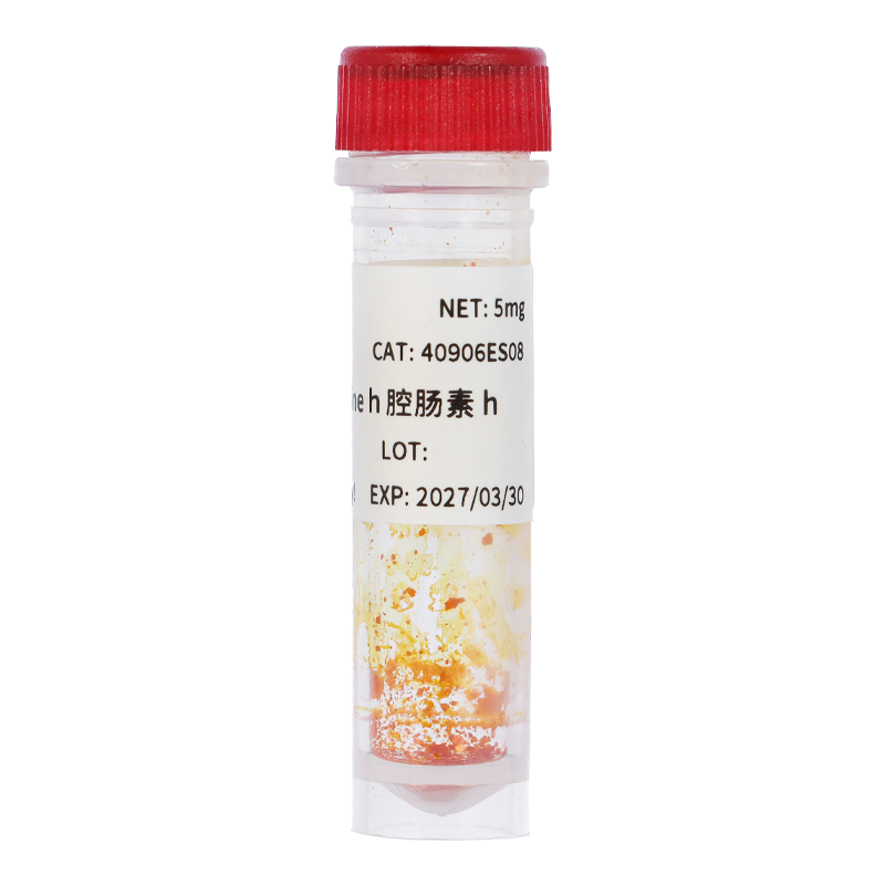 腔肠素h 天然腔肠素去羟基衍生物
40906ES
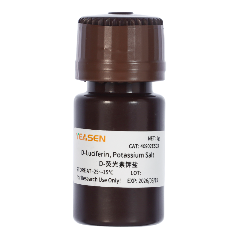 D-荧光素钾盐(D-Luciferin,Potassium Salt...
40902ES
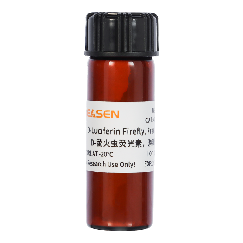 D-萤火虫荧光素游离酸 荧光素酶底物
40903ES
 硫代乙酰胺(Thioacetamide,TAA) 中毒肝炎模型诱导剂
60395ES
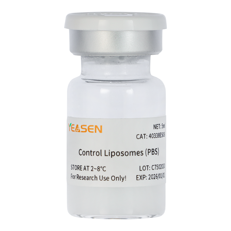 PBS体内巨噬细胞清除剂 PBS空白脂质体对照
40338ES
 4-Nitroquinoline N-oxide 4-硝基喹啉-N-...
60392ES
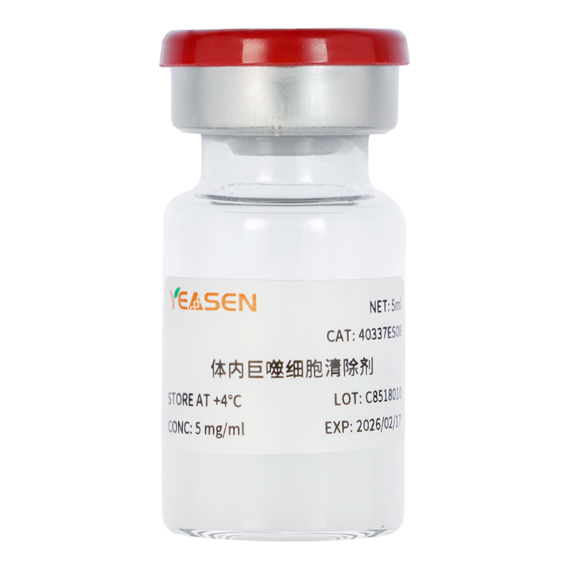 体内巨噬细胞清除剂 Clodronate Liposomes
40337ES
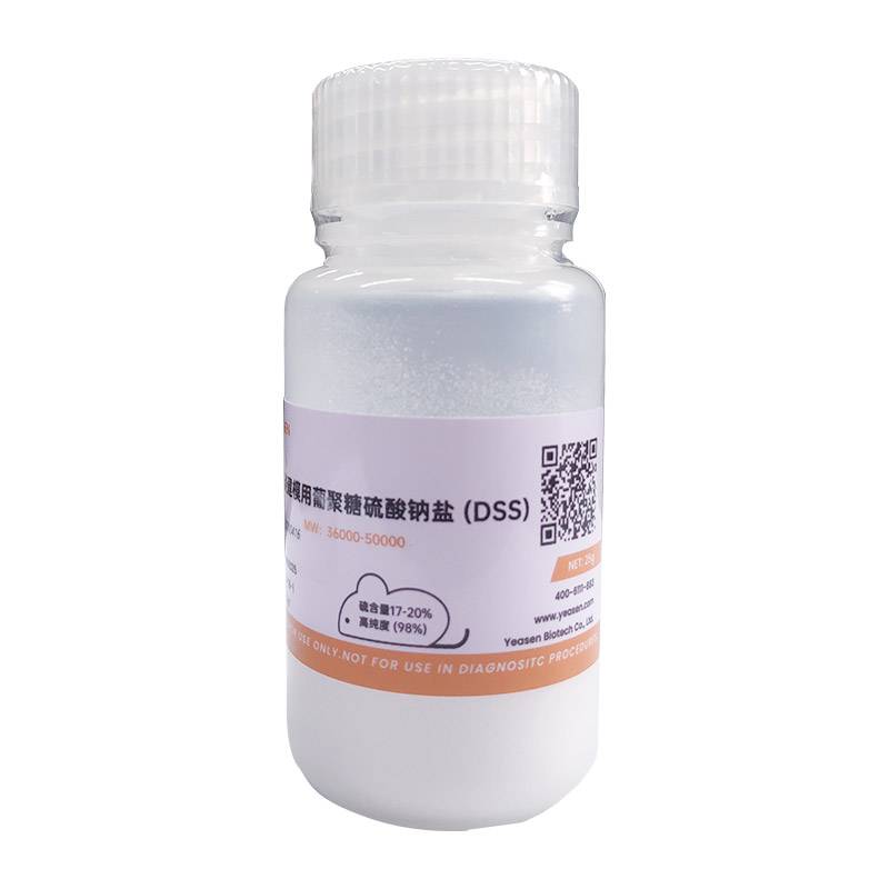 ColitCare®DSS葡聚糖硫酸钠盐(结肠炎建模用) Mw 36...
60316ES
