
Post RFQ
Service Hotline
19920083827
Tick the specs you need to submit them together; or enquire one by one. Cat. numbers are carried into the RFQ.
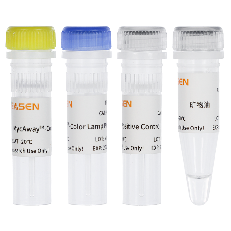
Q:40612 一步法快速支原体检测试剂盒(防污染版)的检测原理?
A:本试剂盒中加入了UDG 酶,以便去除含 dU 的PCR 产物气溶胶污染,在极大程度上抑制假阳性反应,提高检测的准确度。而且加入的 UDG 酶在室温下起作用,且对温度感、易于灭活,不影响实验结果。若细胞培养物被支原体污染,支原体 DNA 的保守序列会被大量、快速地扩增,使反应液由蓝紫色变成天蓝色,结果肉眼可辨,无需电泳。
Q:一步法快速支原体检测试剂盒使用方法(防污染版)?
A:本试剂盒使用更为简便,只需吸取 1μL 疑似支原体污染的细胞培养上清加入反应体系中,先将样品在 25~37 ℃放置 5 min,再在水浴锅或 PCR 仪中,恒温 63 °C 孵育 60 min。观察反应液由蓝紫色变成天蓝色,结果肉眼可辨,无需电泳。
Q:用 40612 一步法支原体检测试剂盒检测 DMSO、乙醇等有机溶剂中是否存在支原体污染,发现实验组均是天蓝色,是不是 DMSO、乙醇等时机中存在支原体污染?
A:支原体试剂盒在乙醇、DMSO 有机溶剂,以及强酸强碱环境中,不稳定易变性,导致颜色变色为天蓝色。这款试剂盒只能用于细胞检测,不可用于有机溶剂、强酸强碱环境下检测。
Q:我检测的是这个里面的液体,加入之后PCR管里的颜色立马变成了天蓝色?是什么原因呢?检测胰酶-EDTA,加入样品之后,直接变色了?
A:40612原理:Mg2++HNB=蓝紫色,随着PCR增加,焦磷酸不断增多,焦磷酸与Mg2+结合,HNB游离,显示天蓝色。这个试剂里面有磷酸二氢钠可能是这个离子与Mg2+结合而立马产生化学反应导致的,EDTA可以螯合钙镁离子,从而导致反应加速发生变色反应。
Q:阳性对照的浓度时多少?
A:10的5次方copies/ul。
Q:阳性对照里的是支原体吗?
A:不是的,是线性化的片段。
Published citations
[1] Shi N, Yang Q, Zhang H, et al. Restoration of dystrophin expression in mice by suppressing a nonsense mutation through the incorporation of unnatural amino acids. Nat Biomed Eng. 2022;6(2):195-206. doi:10.1038/s41551-021-00774-1(IF:25.671)
[2] Zhang S, Yu F, Che A, et al. Neuroendocrine Regulation of Stress-Induced T Cell Dysfunction during Lung Cancer Immunosurveillance via the Kisspeptin/GPR54 Signaling Pathway. Adv Sci (Weinh). 2022;9(13):e2104132. doi:10.1002/advs.202104132(IF:16.806)
[3] Li Y, Xue B, Zhang M, et al. Transcription-coupled structural dynamics of topologically associating domains regulate replication origin efficiency. Genome Biol. 2021;22(1):206. Published 2021 Jul 12. doi:10.1186/s13059-021-02424-w(IF:13.583)
[4] Wu L, Xu Y, Zhao H, et al. FcγRIIB potentiates differentiation of myeloid-derived suppressor cells to mediate tumor immunoescape. Theranostics. 2022;12(2):842-858. Published 2022 Jan 1. doi:10.7150/thno.66575(IF:11.556)
[5] Tan B, Shi X, Zhang J, et al. Inhibition of Rspo-Lgr4 Facilitates Checkpoint Blockade Therapy by Switching Macrophage Polarization. Cancer Res. 2018;78(17):4929-4942. doi:10.1158/0008-5472.CAN-18-0152(IF:9.130)
[6] Yan G, Zhao H, Zhang Q, et al. A RIPK3-PGE2 Circuit Mediates Myeloid-Derived Suppressor Cell-Potentiated Colorectal Carcinogenesis. Cancer Res. 2018;78(19):5586-5599. doi:10.1158/0008-5472.CAN-17-3962(IF:9.130)
[7] Gu Z, Shi C, Li J, et al. Palbociclib-based high-throughput combination drug screening identifies synergistic therapeutic options in HPV-negative head and neck squamous cell carcinoma. BMC Med. 2022;20(1):175. Published 2022 May 12. doi:10.1186/s12916-022-02373-6(IF:8.775)
[8] Wu L, Zhang X, Zheng L, et al. RIPK3 Orchestrates Fatty Acid Metabolism in Tumor-Associated Macrophages and Hepatocarcinogenesis. Cancer Immunol Res. 2020;8(5):710-721. doi:10.1158/2326-6066.CIR-19-0261(IF:8.728)
[9] Qin J, Zhang X, Tan B, et al. Blocking P2X7-Mediated Macrophage Polarization Overcomes Treatment Resistance in Lung Cancer. Cancer Immunol Res. 2020;8(11):1426-1439. doi:10.1158/2326-6066.CIR-20-0123(IF:8.728)
[10] Cao M, Huang W, Chen Y, et al. Chronic restraint stress promotes the mobilization and recruitment of myeloid-derived suppressor cells through β-adrenergic-activated CXCL5-CXCR2-Erk signaling cascades. Int J Cancer. 2021;149(2):460-472. doi:10.1002/ijc.33552(IF:7.396)
[11] Yao R, Alkhawtani AYF, Chen R, Luan J, Xu M. Rapid and efficient in vivo angiogenesis directed by electro-assisted bioprinting of alginate/collagen microspheres with human umbilical vein endothelial cell coating layer [published correction appears in Int J Bioprint. 2020 Sep 17;6(4):309]. Int J Bioprint. 2019;5(2.1):194. Published 2019 Jun 24. doi:10.18063/ijb.v5i2.1.194(IF:6.638)
[12] Liu L, Deng Y, Zheng Z, et al. Hsp90 Inhibitor STA9090 Sensitizes Hepatocellular Carcinoma to Hyperthermia-Induced DNA Damage by Suppressing DNA-PKcs Protein Stability and mRNA Transcription. Mol Cancer Ther. 2021;20(10):1880-1892. doi:10.1158/1535-7163.MCT-21-0215(IF:6.261)
[13] Wu S, Yang X, Tang W, et al. Chemotherapeutic Risk lncRNA-PVT1 SNP Sensitizes Metastatic Colorectal Cancer to FOLFOX Regimen. Front Oncol. 2022;12:808889. Published 2022 Mar 31. doi:10.3389/fonc.2022.808889(IF:6.244)
[14] Wu Q, Xuan YF, Su AL, Bao XB, Miao ZH, Wang YQ. TNKS inhibitors potentiate proliferative inhibition of BET inhibitors via reducing β-Catenin in colorectal cancer cells. Am J Cancer Res. 2022;12(3):1069-1087. Published 2022 Mar 15. (IF:6.166)
[15] Wu L, Zhao KQ, Wang W, et al. Nuclear receptor coactivator 6 promotes HTR-8/SVneo cell invasion and migration by activating NF-κB-mediated MMP9 transcription. Cell Prolif. 2020;53(9):e12876. doi:10.1111/cpr.12876(IF:5.753)
[16] Wei Z, Wang Y, Peng J, et al. CircRFWD3 promotes HNSCC metastasis by modulating miR-27a/b/PPARγ signaling. Cell Death Discov. 2022;8(1):285. Published 2022 Jun 11. doi:10.1038/s41420-022-01066-6(IF:5.241)
[17] Chen W, Weng Z, Xie Z, et al. Sequencing of methylase-accessible regions in integral circular extrachromosomal DNA reveals differences in chromatin structure. Epigenetics Chromatin. 2021;14(1):40. Published 2021 Aug 23. doi:10.1186/s13072-021-00416-5(IF:4.954)
[18] Wu Z, Zheng M, Zhang Y, et al. Hsa_circ_0043278 functions as competitive endogenous RNA to enhance glioblastoma multiforme progression by sponging miR-638. Aging (Albany NY). 2020;12(21):21114-21128. doi:10.18632/aging.103603(IF:4.831)
[19] Wang L, Zhou Y, Cao C, et al. The exon 12-containing LHX6 isoforms promote cervical cancer cell proliferation by regulating the MAPK signaling pathway [published online ahead of print, 2022 Apr 5]. Cancer Med. 2022;10.1002/cam4.4734. doi:10.1002/cam4.4734(IF:4.452)
[20] Zhang Q, Yan G, Lei J, et al. The SP1-12LOX axis promotes chemoresistance and metastasis of ovarian cancer. Mol Med. 2020;26(1):39. Published 2020 May 6. doi:10.1186/s10020-020-00174-2(IF:4.096)
[21] Hu J, Wu Q, Wang Z, et al. Inhibition of CACNA1H attenuates doxorubicin-induced acute cardiotoxicity by affecting endoplasmic reticulum stress. Biomed Pharmacother. 2019;120:109475. doi:10.1016/j.biopha.2019.109475(IF:3.743)
[22] Wang T, Lin F, Sun X, et al. HOXB8 enhances the proliferation and metastasis of colorectal cancer cells by promoting EMT via STAT3 activation. Cancer Cell Int. 2019;19:3. Published 2019 Jan 3. doi:10.1186/s12935-018-0717-6(IF:3.439)
[23] Meng LL, Wang JL, Xu SP, et al. Low serum gastrin associated with ER+ breast cancer development via inactivation of CCKBR/ERK/P65 signaling. BMC Cancer. 2018;18(1):824. Published 2018 Aug 16. doi:10.1186/s12885-018-4717-7(IF:3.288)
[24] Ma J, Liu X, Liu P, et al. Identification of a new p53 responsive element in the promoter region of anillin. Int J Mol Med. 2020;45(5):1563-1570. doi:10.3892/ijmm.2020.4527(IF:3.098)
[25] Chen L, Chen L, Wan L, et al. Matrine improves skeletal muscle atrophy by inhibiting E3 ubiquitin ligases and activating the Akt/mTOR/FoxO3α signaling pathway in C2C12 myotubes and mice. Oncol Rep. 2019;42(2):479-494. doi:10.3892/or.2019.7205(IF:3.041)
[26] Men XM, Xu ZW, Tao X, Deng B, Qi KK. FNDC5 expression closely correlates with muscle fiber types in porcine longissimus dorsi muscle and regulates myosin heavy chains (MyHCs) mRNA expression in C2C12 cells. PeerJ. 2021;9:e11065. Published 2021 Apr 19. doi:10.7717/peerj.11065(IF:2.984)
[27] Zhao YQ, Wu T, Wang LF, et al. Targeting MUC1-C reverses the cisplatin resistance of esophageal squamous cell carcinoma in vitro and in vivo. Transl Cancer Res. 2021;10(2):645-655. doi:10.21037/tcr-20-2495(IF:1.241)
 细胞支原体污染处理新思路——支原体检测、预防、去除一步到位!
前沿资讯
 支原体污染整体解决方案
应用方向
 NAT法|支原体qPCR检测和方法验证
技术讲解
 遇到支原体污染,如何挽救您无比珍贵的细胞?
技术讲解
 生物制品中快速精准检测支原体污染的方法
技术讲解
细胞状态突然变差?支原体污染可能是你忽略的元凶
应用方向
细胞支原体污染如何判断?检测方法与鉴别要点一篇讲透
应用方向
细胞支原体污染清除+预防:一体化解决方案,省时省力
应用方向
2周高效清除细胞支原体污染:从方案到验证,一步不落
应用方向
翌圣生物支原体qPCR检测试剂盒『快速 - 精准 - 合规』
前沿资讯
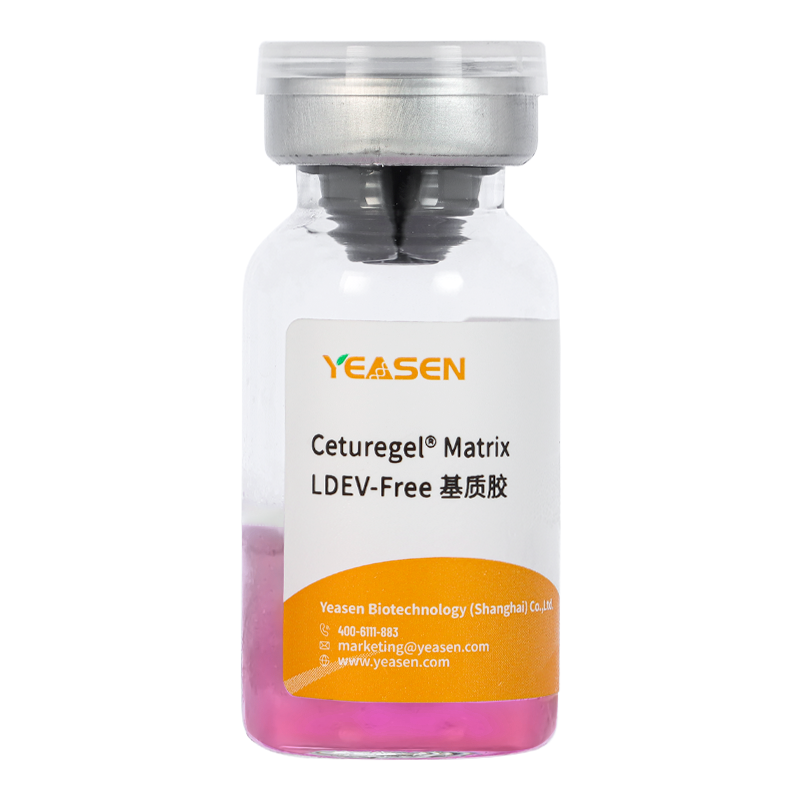 Ceturegel® Matrix LDEV-Free基质胶
40183ES
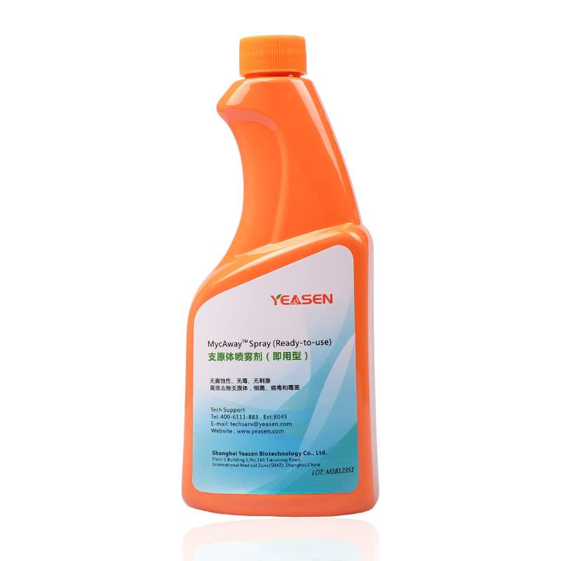 MycAway® Spray (Ready-to-use) 支原体喷...
40605ES
 Ceturegel® Matrix for Organoid cul...
40192ES
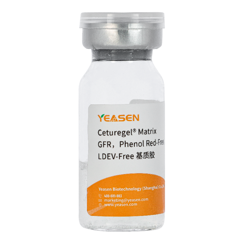 Ceturegel® Matrix GFR,Phenol Red-F...
40186ES
 MycAway® Treatment(1000×)-Mycoplas...
40607ES
 Ceturegel® Matrix High Concentrati...
40187ES
 Lysozyme 溶菌酶
10402ES
 MycAway® Prophylactic (2000×) - My...
40608ES