Welcome to BioReagent Hub Hotline:19920083827
本站自营/精选产品 www.yeasen.com  |  价格与库存以本站实时信息为准
本站精选 · 品质自营查看原始页面 ›
Vendor Tel:400-6111-883
Product data sheet PD-40203ES
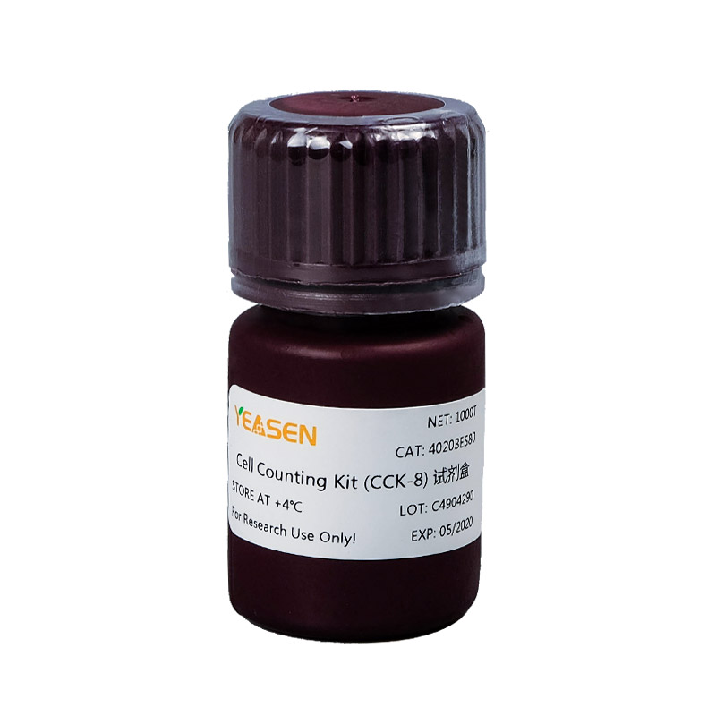
Cell Counting Kit (CCK-8) CCK-8试剂盒 Cell Counting Kit (CCK-8) CCK-8试剂盒
Enquiry

Tell us your volume and application; we reply with a quote.

Start enquiry

Cell Counting Kit (CCK-8) CCK-8试剂盒

Scan for product
酶标仪药物筛选细胞增殖测定细胞毒性测定肿瘤药敏试验生物因子活性检测
Product no.40203ES Sizes5 options Documents1 files SourceYeasen · Official
Cat. No. 40203ES60
现货 $118.00 促$135.00
Qty 1
Enquire now
Enquire / bulk Data sheet Compare

Specifications & Enquiry

Tick the specs you need to submit them together; or enquire one by one. Cat. numbers are carried into the RFQ.
Cat.No Spec Price Status
40203ES60 100 T $118.00促$135.00 现货 Enquire
40203ES76 500 T $398.00促$445.00 现货 Enquire
40203ES80 1000 T $778.00促$875.00 现货 Enquire
40203ES88 3× 1000 T $1,508.00促$1,685.00 现货 Enquire
40203ES92 10× 1000 T $4,388.00促$4,885.00 现货 Enquire
Submit enquiry Price is on application; bulk sizes can be sampled on request.

Overview

Specifications

Brand翌圣生物 Yeasen
Cat.No40203ES
Specification操作简单:产品本身是液体,即开即用;甲臜产物是水溶性的,不需要用有机溶剂溶解。灵敏度高:线性范围广,灵敏度高,重复性好。细胞毒性低:细胞毒性非常低,对后续实验没有影响。稳定性好:培养液中酚红和血清的存在不会对检测造成影响。应用性广:可用于大
Origin翌圣生物科技(上海)股份有限公司
技术平台 酶标仪
应用方法 药物筛选用于筛选药物对细胞增殖或毒性的影响 细胞增殖测定测定细胞增殖能力 细胞毒性测定测定细胞毒性 肿瘤药敏试验用于肿瘤药物敏感性检测 生物因子活性检测检测生物因子对细胞活性的影响
适用物种种属 通用
检测方法 比色法
生物特性参数 适用细胞类型多种哺乳动物细胞系(如293T、GIST-T1、HepG2等)
样本类型 细胞
技术参数 主要成分WST-8 检测波长450nm 储存条件-25~-15℃避光干燥保存 运输条件冰袋运输 有效期2年 操作形式即开即用液体 水溶性产物水溶性,不需要有机溶剂溶解 酚红和血清耐受性酚红和血清的存在不会影响检测 灵敏度高 细胞毒性低
实验类型 细胞增殖检测检测细胞增殖能力 细胞毒性检测检测细胞毒性

FAQ

Q:CCK-8 的保存和稳定性如何?

A:CCK-8 稳定性高,避光条件-20℃有效期 2 年,4℃有效期 1 年。经常使用建议 4℃ 保存,避免反复冻融。CCK 正常颜色为粉红色,若颜色发生明显偏差,质量可能有发生变化。

Q:CCK-8 的细胞毒性如何?

A:CCK-8 毒性非常低,因此 CCK-8 检测完后相同的细胞还能用于其它细胞增殖检测如结晶紫染色法,中性红染色法或 DNA 荧光染色法。

Q:CCK-8 会对活细胞进行染色吗?

A:不会,首先 WST-8 不会进入细胞染色,整个显色反应都是在培养体系中进行的。另外 WST 和 WST-8 甲臜都属于高度水溶性组分,反应结束更换新的培养液,即可清除外源组分。因为 CCK-8 是基于 WST-8 能够被线粒体内的脱氢酶还原生成高度水溶性的橙黄色甲臜产物(Formazan)的原理研制开发的,因而不会对细胞进行染色。

Q:培养基的本底颜色如酚红会不会影响细胞活性的检测呢?

A:不会的,培养基中酚红的吸光度可以在计算时,通过扣除空白孔中本底的吸光度而消去,因此不会对检测造成影响。

Q:在加入 CCK-8 时可否不更换培养基?

A:一般情况下可以不更换培养基。但是如果培养基里有氧化还原性物质的话,有可能会产生误差,必须更换其它培养基。

Q:在实验中OD 值太高或者太低,怎么解决?

A:太高可以缩短加入CCK-8 后的培养时间。例如:可以把加入CCK-8 试剂后的培养时间由 2 小时缩短为 1 小时。此外,可适当减少细胞的数量。太低可以采取 2 个办法: 1、适当增加细胞数量 2、延长加入CCK-8 试剂后的反应时间。

Q:哪些物质会影响 CCK-8 的实验测定结果?

A:当培养体系内含有还原性物质存在时会还原 WST-8,从而使 OD 值增加;当有氧化性物质存在时会抑制还原反应进而使 OD 值减小。

Q:做细胞毒性实验,我的细胞都死了,为什么我加入CCK8试剂后检测的OD值和对照组OD值差不多?

A:①细胞凋亡或者死亡之后乳酸脱氢酶被释放到细胞培养液中,可以长期存在,从而导致检测的OD值偏大。再次说明细胞的活性和OD值并不能完全划等号。建议客户在加入CCK8的时候更换新的培养基,确保得到准确的结果。

②加入细胞中的检测药物是还原性的,从而导致OD值偏高。建议加入CCK8的时候更换新的培养基。

Q:对照组(培养基+CCK8)的值很高,是为什么呢?

A:可以测一下培养基是否含有氧化还原物质,可能是培养基中的物质引起的。

Q:  细胞不贴壁可以测活性吗?预培养是必须的吗?

A: 需要预培养,如果要向保持细胞的最好状态,建议预培养细胞。如果不做细胞预培养 ,细胞内的脱氢酶可能会不稳定。若不做细胞预培养,需在做标准曲线和检测时需要统一检测条件。

Q: CCK-8能否检测细菌细胞?

A: 可以检测大肠杆菌,但不能检测酵母细胞。向100μl E.coli培养液中加入10μl CCK-8溶液,并培养1-4个小时或过夜。

Documents & Citations

Published citations

  1. Chen Z, Jiang L, Liu X, et al. NirFAP680: a highly bright and stable large Stokes shift near-infrared fluorogen-activating protein. Nat Methods. Published online April 21, 2026. doi:10.1038/s41592-026-03061-6 (IF:32.1)
  2. Qian X, Yi W, Yan W, et al. Cryo-Shocked Tumor-Reprogrammed Sonosensitive Antigen-Presenting Cells Improving Sonoimmunotherapy via T Cells and NK Cells Immunity. Adv Mater. Published online February 16, 2025. doi:10.1002/adma.202413289 (IF:27.4)
  3. Yao C, Ni Z, Gong C, et al. Rocaglamide enhances NK cell-mediated killing of non-small cell lung cancer cells by inhibiting autophagy[J]. Autophagy, 2018, 14(10): 1831-1844. IF(11.1)
  4. X Fan, H Cheng, et al. Incorporation of Polycaprolactone to Cyclodextrin-Based Nanocarrier for Potent Gene Delivery. Macromolecular materials and engineering, Volume303, Issue9 ,September 2018.IF(2.777)
  5. Cheng H, Fan X, et al. Cyclodextrin-Based Star-Like Amphiphilic Cationic Polymer as a Potential Pharmaceutical Carrier in Macrophages.  Macromol Rapid Commun. 2018 May 28. IF(4.441)
  6. Ma Q, Zhang Y, Liang H, et al. EMP3, which is regulated by miR-663a, suppresses gallbladder cancer progression via interference with the MAPK/ERK pathway[J]. Cancer letters, 2018, 430: 97-108.IF(6.491)
  7. Liu Y, Pan Y F, Xue Y, et al. uPAR promotes tumor-like biologic behaviors of fibroblast-like synoviocytes through PI3K/Akt signaling pathway in patients with rheumatoid arthritis[J]. Cellular & molecular immunology, 2018, 15(2): 171. IF(7.551)
  8. Shen, C.T., et al., Metformin reduces glycometabolism of papillary thyroid carcinoma in vitro and in vivo. J Mol Endocrinol, 2017. 58(1): p. 15-23.IF(3.297)
  9. Jiang, J.Z., et al., Metabolic-induced cytotoxicity of diosbulbin B in CYP3A4-expressing cells. Toxicol In Vitro, 2017. 38: p. 59-66.IF(3.105)
  10. Du Z, et al., MicroRNA31-NDRG3 regulation axes are essential for hepatocellular carcinoma survival and drug resistance. Cancer Biomark, 2017.IF(2.392)
  11. Li, X., et al., Placental growth factor silencing ameliorates liver fibrosis and angiogenesis and inhibits activation of hepatic stellate cells in a murine model of chronic liver disease. J Cell Mol Med, 2017.(4.302)
  12. Liu, S., et al., Macrophage infiltration of electrospun polyester fibers. Biomater Sci, 2017.IF(5.831)
  13. Shi, G., et al., Sox9 facilitates proliferation, differentiation and lipogenesis in primary cultured human sebocytes. J Dermatol Sci, 2017. 85(1): p. 44-50.IF(3.675)
  14. Liu, Y., et al., uPAR promotes tumor-like biologic behaviors of fibroblast-like synoviocytes through PI3K/Akt signaling pathway in patients with rheumatoid arthritis. Cell Mol Immunol, 2017.IF(7.551)
  15. Zhang, S., J. Yin and J. Zhong, Chaetocin reactivates the lytic replication of Epstein-Barr virus from latency via reactive oxygen species. Sci China Life Sci, 2017. 60(1): p. 66-71.IF(3.085)
  16. Yang, Q.L., et al., Sweroside ameliorates alpha-naphthylisothiocyanate-induced cholestatic liver injury in mice by regulating bile acids and suppressing pro-inflammatory responses. Acta Pharmacol Sin, 2016. 37(9): p. 1218-28.IF(3.562)
  17. Wei, W.J., et al., Propranolol sensitizes thyroid cancer cells to cytotoxic effect of vemurafenib. Oncol Rep, 2016. 36(3): p. 1576-84.IF(2.976)
  18. Yang, Y., et al., Human CIK Cells Loaded with Au Nanorods as a Theranostic Platform for Targeted Photoacoustic Imaging and Enhanced Immunotherapy and Photothermal Therapy. Nanoscale Res Lett, 2016. 11(1): p. 285.IF(3.125)
  19. Liu, T., et al., Slowly Delivered Icariin/Allogeneic Bone Marrow-Derived Mesenchymal Stem Cells to Promote the Healing of Calvarial Critical-Size Bone Defects. Stem Cells Int, 2016. 2016: p. 1416047.IF(3.989)
  20. Zhou, Y., et al., Synergistic anti-liver fibrosis actions of total astragalus saponins and glycyrrhizic acid via TGF-beta1/Smads signaling pathway modulation. J Ethnopharmacol, 2016. 190: p. 83-90.IF(3.115)
  21. Li, W., et al., MicroRNA-495 regulates starvation-induced autophagy by targeting ATG3. FEBS Lett, 2016. 590(6): p. 726-38.IF(2.999)
  22. Yang, F., et al., Waltonitone inhibits proliferation of hepatoma cells and tumorigenesis via FXR-miR-22-CCNA2 signaling pathway. Oncotarget, 2016. 7(46): p. 75165-75175.IF(2.656)
  23. Li, S., et al., DNA Cleavage and Condensation Activities of Mono- and Binuclear Hybrid Complexes and Regulation by Graphene Oxide. Molecules, 2016. 21(7).IF(3.098)
  24. Li, Z.T., et al., Outer membrane vesicles isolated from two clinical Acinetobacter baumannii strains exhibit different toxicity and proteome characteristics. Microb Pathog, 2015. 81: p. 46-52.IF(2.332)
  25. Xiao, K., et al., Inactivation of BLU is associated with methylation of Sp1-binding site of BLU promoter in gastric cancer. Int J Oncol, 2015. 47(2): p. 621-31.IF(3.333)
  26. Sun, Y.P., et al., Synovium fragment-derived cells exhibit characteristics similar to those of dissociated multipotent cells in synovial fluid of the temporomandibular joint. PLoS One, 2014. 9(7): p. e101896.IF(2.766)
  27. Qiao, N., et al., Haemolytic activity and adjuvant effect of soyasaponins and some of their derivatives on the immune responses to ovalbumin in mice. Int Immunopharmacol, 2014. 18(2): p. 333-9.IF(3.118)

Recommended reading

Related products

✉Email 💬Message 👤Member
↑Top