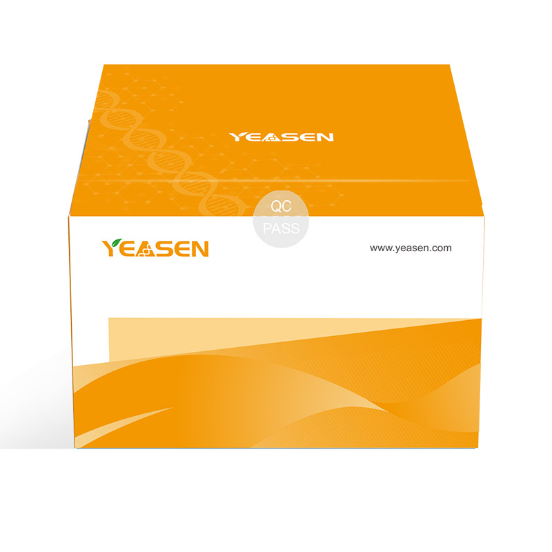
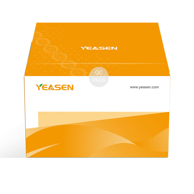
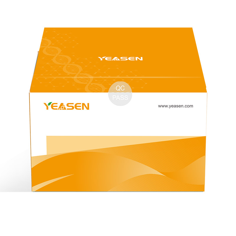
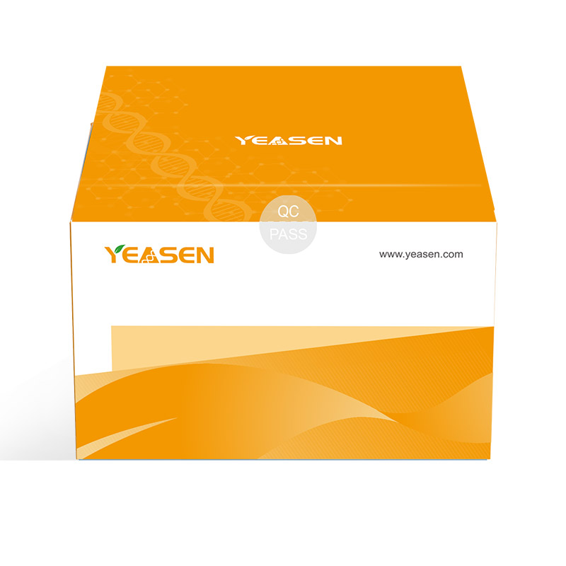
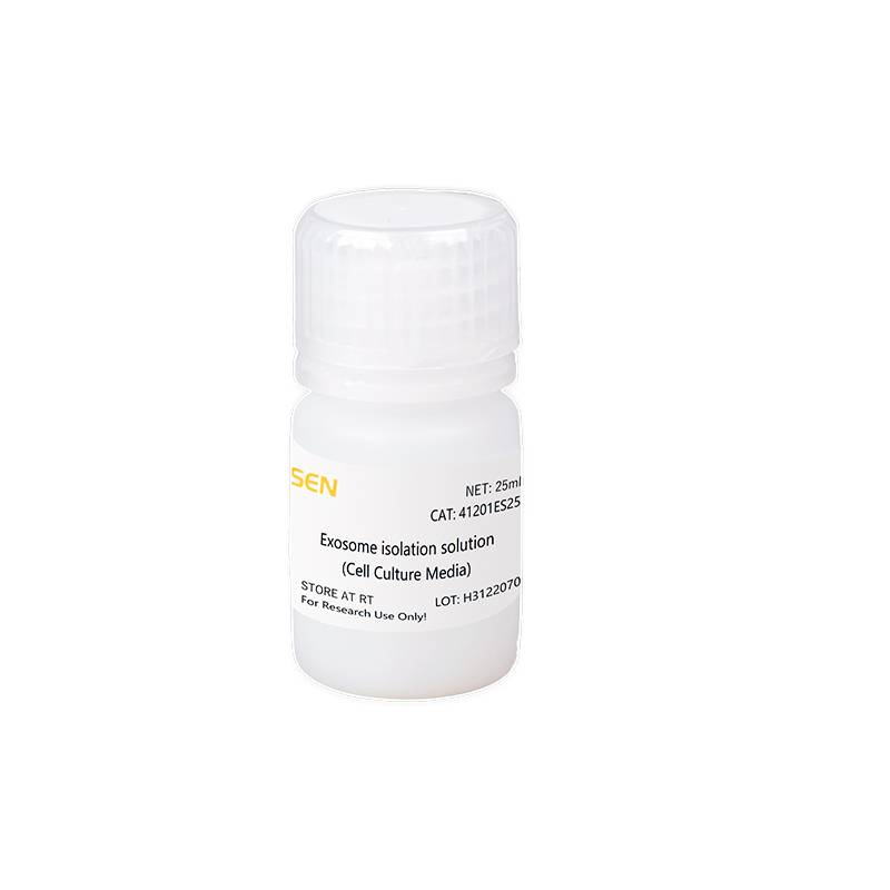
Hieff® Quick exosome isolation kit(for Cell Culture Media) 细胞培养上清外泌体快速抽提试剂
勾选需要的规格可一次提交;也可以逐个询价。货号会一并带入询价单。

Q:外泌体洗涤那步为什么要保留上清液?
A:因为外泌体溶解到上清中了,这个转速下外泌体还是在上清中。
Q:是利用的磁珠法还是柱法?
A:这个不属于纯化,是生物试剂 PEG。
Q:最后得到外泌体沉淀了,为什么还要再洗涤弃沉淀,沉淀是什么?
A:去除提取的或试剂中的不溶性沉淀。
Q4:无外泌体血清培养基(或者无血清培养基)在什么时候使用?
A:细胞在正常含血清的培养基中培养一定的时间后,细胞融合度约为60%-70%时,移去原有含血清的培养基,换成新鲜的无外泌体血清培养基(或者无血清培养基),继续培养24-48h,细胞融合度达到80%-95%左右时收取上清,该上清液即可用于提取外泌体。
Q5:细胞培养过程中的死细胞是否会影响外泌体的提取?
A:会的。在收获细胞时,应确定死亡细胞占比不超过5%。细胞凋亡/死亡过程中会释放大量大小不等的囊泡,它们在外泌体的提取纯化过程中会污染活细胞产生的外泌体,同时有可能产生大量杂质。
Q6:加了抽提试剂离心之后无沉淀,这个现象正常吗?
A:由于某些细胞(如悬浮细胞、干细胞、神经细胞等)中外泌体含量比较低,加了试剂A离心之后肉眼可能无法观察到沉淀,在重悬时使用PBS液朝离心管离心外侧的内壁反复吹打洗脱即可(避免剧烈吹打)。针对提取后的外泌体先进行NTA粒径检测或BCA蛋白定量检测,再决定是否进行后继实验。
Q7:客户外泌体染色之后,是否可以用我们的细胞上清外泌体抽提试剂盒再次提取?对提取的外泌体有没有上限?添加是否也是按照体积比进行添加抽提试剂?
A: 可以的,对重新提取的外泌体没有上限要求,等比例添加就好。也推荐超滤管的方式。
Q8:沉淀法应该是不会对已经染色的外泌体有影响吧?
A: 沉淀法对染色的外泌体没什么影响。
Q9: 细胞培养上清外泌体快速抽提试剂盒,开封之后2-3个月,客户封口保存了,但是现在变成粉末了。可以复溶之后继续使用吗?
A: 可能原因是瓶子没拧紧,不建议再使用了,建议重新购买。
已发表文献
[1] Li Z, Qin X, Bian W, et al. Exosomal lncRNA ZFAS1 regulates esophageal squamous cell carcinoma cell proliferation, invasion, migration and apoptosis via microRNA-124/STAT3 axis. J Exp Clin Cancer Res. 2019;38(1):477. Published 2019 Nov 27. doi:10.1186/s13046-019-1473-8(IF:11.161)
[2] Guan X, Zong ZH, Liu Y, Chen S, Wang LL, Zhao Y. circPUM1 Promotes Tumorigenesis and Progression of Ovarian Cancer by Sponging miR-615-5p and miR-6753-5p. Mol Ther Nucleic Acids. 2019;18:882-892. doi:10.1016/j.omtn.2019.09.032(IF:8.886)
[3] Chen G, Yue A, Wang M, Ruan Z, Zhu L. The Exosomal lncRNA KLF3-AS1 From Ischemic Cardiomyocytes Mediates IGF-1 Secretion by MSCs to Rescue Myocardial Ischemia-Reperfusion Injury. Front Cardiovasc Med. 2021;8:671610. Published 2021 Sep 21. doi:10.3389/fcvm.2021.671610(IF:6.050)
[4] Zhang H, Wang J, Wang Y, et al. Long Non-Coding LEF1-AS1 Sponge miR-5100 Regulates Apoptosis and Autophagy in Gastric Cancer Cells via the miR-5100/DEK/AMPK-mTOR Axis. Int J Mol Sci. 2022;23(9):4787. Published 2022 Apr 26. doi:10.3390/ijms23094787(IF:5.924)
[5] Li F, Niu R, Gao S, et al. Pro-Angiogenesis Role of LINC00662 From Esophageal Squamous Cell Carcinoma Cells-Derived Extracellular Vehicles. Front Bioeng Biotechnol. 2022;10:772514. Published 2022 Apr 1. doi:10.3389/fbioe.2022.772514(IF:5.890)
[6] Luo X, Wei J, Yang FL, et al. Exosomal lncRNA HNF1A-AS1 affects cisplatin resistance in cervical cancer cells through regulating microRNA-34b/TUFT1 axis. Cancer Cell Int. 2019;19:323. Published 2019 Dec 3. doi:10.1186/s12935-019-1042-4(IF:5.722)
[7] Wang ZF, Liao F, Wu H, Dai J. Glioma stem cells-derived exosomal miR-26a promotes angiogenesis of microvessel endothelial cells in glioma. J Exp Clin Cancer Res. 2019;38(1):201. Published 2019 May 17. doi:10.1186/s13046-019-1181-4(IF:5.646)
[8] Chen G, Wang M, Ruan Z, Zhu L, Tang C. Mesenchymal stem cell-derived exosomal miR-143-3p suppresses myocardial ischemia-reperfusion injury by regulating autophagy. Life Sci. 2021;280:119742. doi:10.1016/j.lfs.2021.119742(IF:5.037)
[9] Shang A, Wang X, Gu C, et al. Exosomal miR-183-5p promotes angiogenesis in colorectal cancer by regulation of FOXO1. Aging (Albany NY). 2020;12(9):8352-8371. doi:10.18632/aging.103145(IF:4.831)
[10] Ma R, Liang Z, Shi X, et al. Exosomal miR-486-5p derived from human placental microvascular endothelial cells regulates proliferation and invasion of trophoblasts via targeting IGF1. Hum Cell. 2021;34(5):1310-1323. doi:10.1007/s13577-021-00543-x(IF:4.174)
[11] Hu H, Xu H, Lu F, et al. Exosome-Derived miR-486-5p Regulates Cell Cycle, Proliferation and Metastasis in Lung Adenocarcinoma via Targeting NEK2 [retracted in: Front Bioeng Biotechnol. 2021 Mar 26;9:681918]. Front Bioeng Biotechnol. 2020;8:259. Published 2020 Apr 8. doi:10.3389/fbioe.2020.00259(IF:3.644)
[12] Sun LP, Xu K, Cui J, et al. Cancer‑associated fibroblast‑derived exosomal miR‑382‑5p promotes the migration and invasion of oral squamous cell carcinoma. Oncol Rep. 2019;42(4):1319-1328. doi:10.3892/or.2019.7255(IF:3.041)
[13] Liu Y, Fu W, Cao X, et al. Delivery of miR-224-5p by Exosomes from Cancer-Associated Fibroblasts Potentiates Progression of Clear Cell Renal Cell Carcinoma. Comput Math Methods Med. 2021;2021:5517747. Published 2021 May 24. doi:10.1155/2021/5517747(IF:2.238)
[14] Song H, Zhang X, Chen R, et al. Cortical Neuron-Derived Exosomal MicroRNA-181c-3p Inhibits Neuroinflammation by Downregulating CXCL1 in Astrocytes of a Rat Model with Ischemic Brain Injury. Neuroimmunomodulation. 2019;26(5):217-233. doi:10.1159/000502694(IF:1.351)
 如何远离外泌体研究烦恼(内附解决方案)
应用方向
 干货分享|想了解外泌体在疾病诊断与治疗上的应用方法?看这篇就够了!
应用方向
 外泌体抑制剂——GW4869
应用方向
 国自然热点 | Yeasen提供外泌体系列解决方案
应用方向
 外泌体核酸提取的原理及应用
应用方向
 外泌体研究历程,你知多少?
技术讲解
 外泌体(exosome)研究整体思路-标记、分离、提取、鉴定
应用方向
 吐血整理外泌体标记大法(附原文献)
文献解读